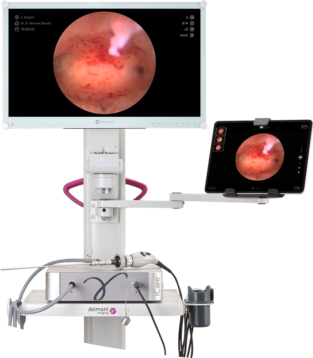

Hysteroscopy, a direct visualization of the uterine cavity
Hysteroscopy is a minimally invasive medical imaging technique used to examine the inside of the uterus, the cervical canal, and the fallopian tube openings. Performed vaginally, it involves inserting a hysteroscope—a thin optical tube equipped with a camera, a light source, and an irrigation system—through the cervix to obtain a direct and precise view of the uterine cavity. There are two types of hysteroscopy: diagnostic hysteroscopy, used to investigate the cause of gynecological problems (abnormal bleeding, infertility, polyps, etc.), and operative hysteroscopy, which is used to treat certain abnormalities (removal of polyps, fibroids, septa, etc.). This procedure is essential, both for establishing an accurate diagnosis and for effective management of uterine pathologies, while preserving the patient's comfort as much as possible.

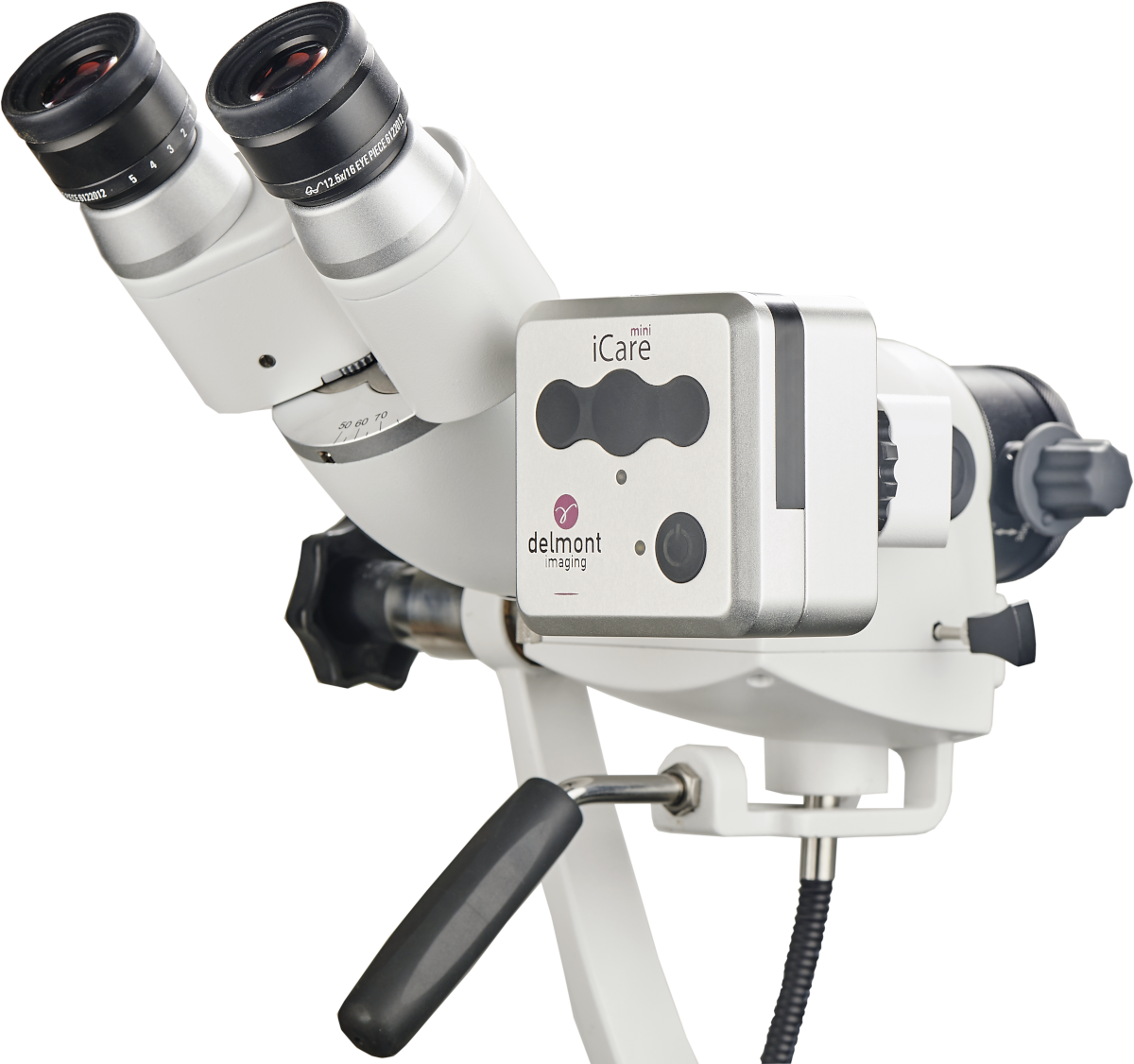
Colposcopy, an examination of the cervix
Colposcopy, an examination of the cervix. Colposcopy is a non-invasive gynecological examination that allows for a detailed observation of the cervix, vagina, and vulva using a colposcope. This device, equipped with a magnifying optical system and a light source, provides precise visualization of the tissues without direct contact. The examination is generally recommended after an abnormal Pap smear or the detection of HPV (human papillomavirus), the main risk factor for precancerous lesions of the cervix. Dyes such as acetic acid or Lugol's solution may be applied to better visualize suspicious areas and, if necessary, to perform targeted biopsies. Beyond diagnosis, colposcopy can also be used to support certain therapeutic procedures, such as conization, which involves removing a portion of the cervix in cases of confirmed lesions. This examination therefore plays a key role in the early detection and management of cervical pathologies, while remaining a simple, quick and well-tolerated procedure.